
Pneumonia
Introduction
Pneumonia is an infection of the lung tissue. The alveoli become filled with micro-organisms, fluid and inflammatory cells causing impaired gaseous exchange and consolidation on Chest X-ray.
Pneumonia has many potential causes. Both viral and bacterial. Below are the most common causes.
Strep pneumoniae (commonest), H. influenza (more common in older, COPD, Chronic lung conditions), Staph aureus (post influenza, IVDU)
Atypical Bacteria – Mycoplasma pneumoniae (younger), C. pneumoniae, Legionella spp (commoner in smokers, outbreaks due to contaminated water)
Viruses – COVID 19, Influenza, RSV,
Hospital Acquired – E.coli, Klebsiella pneumoniae, Enterobacter spp, Pseudomonas, S. aureus, MRSA
Aspiration Pneumonia – Usual bacteria & Gut flora (anaerobes, enteric gram -ves)
Treatment for pneumonia varies and depends on
Community Acquired versus Hospital Acquired (in patient > 48 hrs/recently discharged) versus Aspiration
How unwell the patient is. (CURB 65)
Risk Factors e.g. immunosuppression, Asthma/COPD, Alcoholism, Chronic conditions, risk of aspiration
Clinical Features
Symptoms
Systemic
Chills, fevers, rigors, anorexia, lethargy
Chest
Cough, change in colour or sputum if chronic cough, dyspnoea, pleuritic chest pain
Signs
Signs of sepsis
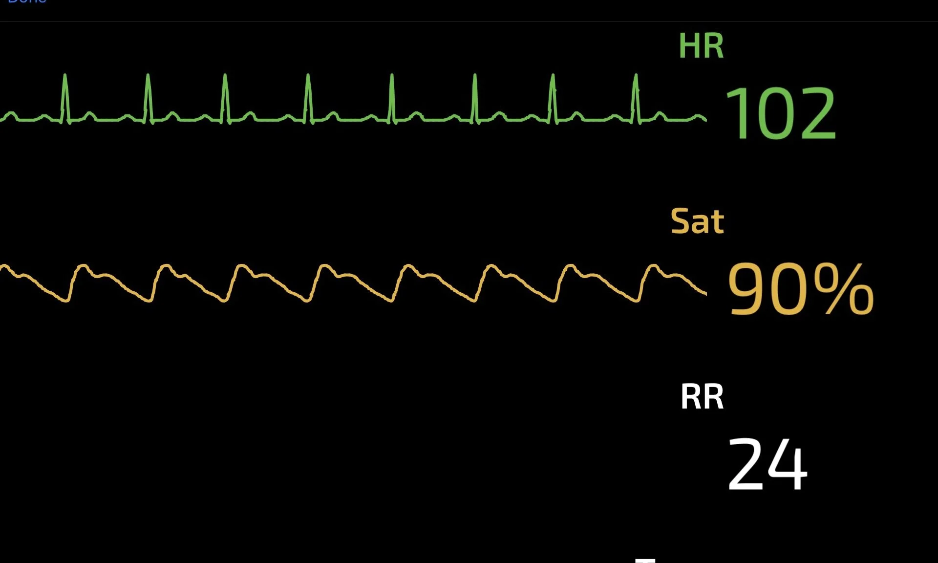
fever, tachycardia, hypotension, hypoxia, tachypnoea
Chest Signs
Focal Decreased air entry, crackles, bronchial breath sounds, dullness to percussion, increased focal resonance
Complications
Respiratory Complications
Pleural effusion, empyema, Cavitation, Abscess Formation
Systemic Complications
Bacteraemia, Septic Shock + Multiorgan Failure, Respiratory Failure
Differential Diagnosis
Respiratory
LRTI - bacterial or viral
Bronchiectasis
Bronchitis
Asthma/COPD exacerbation
PE
TB
Malignancy
ARDS
Cardiac
CCF
Angina
ACS
Pericarditis
Myocarditis
Infective Endocarditis
Biliary Pathology
Cholecystitis
Choledocholithiasis
Clinical Investigation

Bedside
Arterial Blood Gas (if Hypoxic)
Low pH = acidosis (resp or metabolic i.e. sepsis)
PaO2 < 8kPA = Respiratory Failure.
with normal or low PaCO2 = Type 1 resp failure
with high PaCO2 (>6.7kPa) = Type 2 resp failure
High PaCO2 = Resp Acidosis. Low PaCO2 = Resp Alkalosis (Tachypnoea)
Electrolytes, Hb, Lactate, BM
ECG
? Cardiac cause for symptoms
POCUS
Lung US more sensitive and specific for consolidation than CXR
Bedside Echo - ? CCF, ? pericardial effusion

Laboratory
Bloods Tests
Inflammatory markers - High neutrophils, Low lymphocytes (viral), High CRP
U&E – AKI, Low Na often associated with chest sepsis
LFT – often abnormal, especially with Mycoplasma
Mycoplasma serology
Blood cultures if ? sepsis
Sputum for Culture
Urine for Culture and Legionella/Pneumococcal Antigen
Nasal/Throat Swab for Viral PCR

Radiology
CXR
Consolidation, air bronchograms, parapneumonic effusion, Cavitation
Normal CXR doesn’t out rule pneumonia as CXR changes can lag behind clinical findings
CT Chest
Diagnosis may be made when looking for other pathology e.g. PE on CTPA. May have role in atypical pneumonia e.g. COVID 19
CURB65 Score
CURB 65 grades severity of pneumonia and likely mortality rates
5 criteria. 1 point each. Score 0-5
1 = 1% Mortality (mild pneumonia) 2 = 8%, 3 =20%, 4 = 40%, 5 = 60%
C Confusion
U Urea > 7
R RR > 30
B BP. Systolic <90mmHg or Diastolic < 60mmHg
65 Age > 65 years
Management and Disposition
Initial Resuscitation
***PPE and appropriate isolation if ? COVID
Resuscitation as required. ABC approach
Oxygen as required to keep sats > 94%, or > 88% in COPD
Assess for volume depletion and IV fluids as required.
May require vasopressors if septic and non responsive to fluid challenge
Symptomatic Treatment
Analgesia as required
Encourage rest, fluids and not to smoke
Nebulised bronchodilators e.g. salbutamol, if associated bronchospasm
Specific Treatment
Antibiotics as per local guidelines. Below is an example of SJH guidelines
Mild CAP
Amoxicillin 500mg TDS PO x 5/7
Mod/Severe CAP (includes atypical cover)
Co-Amoxyclav 1.2 g TDS IV + Clarithromycin 500mg BD PO/IV
Hospital Acquired Pneumonia (HAP)
Piperacillin-Tazobactam 4.5g QDS IV +/- IV Amikacin +/- IV Vancomycin (if hx of MRSA)
Disposition
Mild Cases (Curb 0 -1) can be managed in the community with PO antibiotics
Moderate (Curb 2) and Severe (Curb 3-5) need to be managed in the hospital setting with IV antibiotics and supportive care +/- ventilatory and organ support in the ICU setting if appropriate
References
1. NICE Guidance. Pneumonia in adults. Quality Standard [QS110] Jan 2016
2. File TM et al. Epidemiology, pathogenesis and microbiology of community-acquired pneumonia in adults. Uptodate.com
3. Putland M, Cameron P et al. Chapter 6.3 Community Acquired Pneumonia. The Textbook of Adult Emergency Medicine 4th Edition
4. British Thoracic Society community acquired pneumonia guideline. Oct 2009
5. SJH Prescriber Capsule. Empiric Antimicrobial Guidelines.