

Transient Ischaemic Attack (TIA)
Introduction
TIA or transient ischaemic attack is defined as a transient episode of neurologic dysfunction caused by focal brain, spinal cord or retinal ischemia without acute infarction. TIA is a common presentation to the emergency department.
It is very important to diagnose TIA and investigate the underlying pathology. The weakness is transient and resolves completely but TIA substantially increases the future risk of having an ischaemic stroke. On average, the annual risk of future ischaemic stroke after a TIA is 3–4%, with an incidence as high as 11% over the next 7 days and 24–29% over the following 5 years.
As the name suggests the final pathological pathway for a TIA is ischaemia. There can be two possible pathways leading to this.
1. Atherosclerosis
2. Embolism
Risk Factors for TIA
Atherosclerotic risk factors
Age >65, Diabetes, HTN, Hyperlipidemia, Smoking, Obesity, Hypertension
Embolic risk factors
Atrial fibrillation, undiagnosed PFO, valvular heart diseases, long bone fractures
Clinical Features
The clinical features of TIA are essentially same as that of an ischaemic stroke. The only differentiating feature is a complete resolution of symptoms and no sign of ischaemia on imaging. Clinical features depend on the vascular territory of the brain involved.
History taking is the key to diagnosing a TIA. A TIA may last only minutes, and symptoms often resolve before the patient presents to a clinician. Thus, historical questions should be addressed not just to the patient but also to anyone who might have witnessed the event.
A thorough physical exam is imperative. Symptoms may have resolved from the patient’s perspective but there might be subtle clinical signs which may lead to diagnosis of an ischaemic stroke rather than a TIA. The goals of the physical examination are to uncover any neurologic deficits, to evaluate for underlying cardiovascular risk factors and to seek any potential thrombotic or embolic source of the event.
Anterior Circulation TIAs
70% of TIAs
Anterior circulation originates from the internal carotid artery and generally supplies the anterior two thirds of the cerebral hemispheres including the basal ganglia
Ischaemia effecting this area generally presents with
Hemiparesis, hemianopia, dysarthria, alexia, agraphia, facial droop, neglect, aphasia etc
Posterior Circulation TIAs
25-30% of TIAs
Posterior circulation of the brain originates from the vertebral artery
Transient occlusion of this system can result in
Cerebellar signs, vertigo, nausea, vomiting, diplopia
Investigations

Bedside
ECG - To look for arrhythmias particularly atrial fibrillation which is the most common embolic arrhythmia.
Blood sugar - Hypoglycemia is one of the most common stroke / TIA mimics
VBG – Gives a rapid assessment of electrolyte abnormalities and haemoglobin. Disturbance in either can mimic a TIA.

Laboratory
FBC – to look for anaemia, thrombocytosis, raised WBCs.
Urea & Electrolytes – to look for hyponatremia, hypokalemia and other electrolyte abnormalities.
CRP - elevated inflammatory markers could raise suspicion for infection which can mimic TIA/stroke
Fasting lipids, Fasting sugars and HbA1c will need to be done by the medical service that are following up the patient as part of their TIA work up. Younger patients may require work up for clotting disorders. These are not routine ED tests.
Radiology
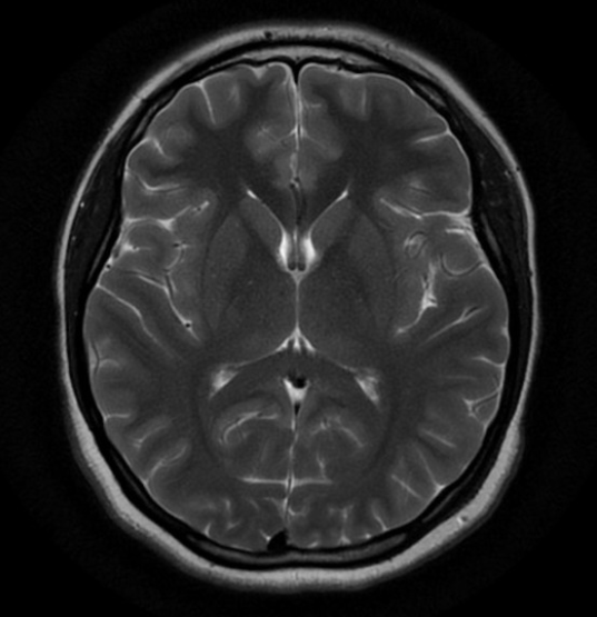
All patients who present with TIA should have neuroimaging (CT / MRI Brain) done within 24 hours of presentation to the emergency department. The aim of imaging is to look for any areas of infarction and to rule out other pathology e.g. bleeds, space occupying lesion.
NICE recommend MRI Brain with diffusion weighted images as first line and advocate against CT brain (unless an alternate pathology is more likely than TIA).
As part of their TIA work up they should have early in patient or outpatient
Carotid Dopplers
Transthoracic echo
Management & Dispostion
Initial Resuscitation
A true TIA patient would rarely need resuscitation. If it is needed you must consider alternate diagnoses
The main differential with a TIA is an acute Ischaemic stroke. As soon as diagnosis of acute stroke is considered “Stroke Call” should be initiated.
The next priority is to exclude stroke mimics such as hypoglycaemia
Specific Treatment
Once neuro-imaging is normal = Aspirin 300mg PO
When diagnosis is made the aim is to seek and treat the potential thrombo-embolic source.
Carotid Dopplers
Echo +/- bubble study if young
Fasting Bloods
Coagulation screen if young
Disposition
These patients are at a very high risk (as high as 11% in next 7 days) of have an ischaemic stroke. The idea is to focus on prevention of this by performing full TIA work up
Should be seen by a stroke specialist within 24 hours
either as an in-patient
or if otherwise well and no confounding factors in a rapid access TIA clinic
References
NICE Guideline. Stroke and transient ischaemic attack in over 16s: diagnosis and initial management. May 2019
This blog was written by Dr Mustafa Mehmood and was last updated in Feb 2021