
Bowel Obstruction
Introduction
Bowel obstruction is generally described as
Small Bowel (80%) or Large Bowel (20%)
Complete or partial
Simple or strangulated
Causes
Small Bowel Obstruction
Adhesions from previous surgery/infection (~75% of cases)
External Hernias – inguinal, femoral, incisional, umbilical
Neoplasms
Rarer causes = strictures e.g. IBD, internal hernias, foreign bodies
Paediatrics = Intussusception, malrotation, volvulus, congenital lesions
Large Bowel Obstruction
Neoplasm
Diverticulitis
Volvulus - Caecal or sigmoid.
Sigmoid more common in elderly, chronically constipated, neuro conditions
Rarer = Hernias, Strictures, IBD, Extra intestinal tumours, Faecal impaction
Clinical Features
Symptoms
Pain
Initially colicky. Can be more severe and constant with strangulation
Vomiting
Occurs more commonly and earlier in SBO. Bilious vomiting = proximal obstruction
Occurs later in LBO. Faeculant vomiting implies distal obstruction
Constipation/Not passing flatus
Late sign. Passing stool or flatus does NOT out rule diagnosis
Bloating
Signs
Signs of sepsis - concerning for perforation
Signs of dehydration
Abdominal Signs
Scars, Hernias, Distension (more common with LBO),
Tenderness. If frank rebound or rigidity concerning for peritonitis
Hypertympanic percussion note (more common in LBO)
Bowel Sounds – may initially be increased but over time become silent
Complications
Volume depletion from vomiting and third space losses
Electrolyte disturbances
Hypokalaemia
Hyponatraemia
Hypernatraemia
metabolic alkalosis from vomiting
Lactic acidosis from ischaemia/sepsis
Strangulation of bowel
ischaemia
perforation & peritonitis
septicaemia
shock
death
Differential Diagnosis
Gastro-intestinal
Diverticulitis
Appendicitis
Psuedo-obstruction
Inflammatory bowel disease
Ileus
Vascular
Mesenteric ischaemic
Ruptured AAA
Infective / Inflammatory
Cholecystitis
Pancreatitis
Pelvic inflammatory disease (PID)
Metabolic
Diabetic ketoacidosis (DKA)
Hyperosmolar hyperglycaemic state (HHS)
Clinical Investigations
Bedside
Urinalysis – can aid in excluding UTI. Urinary BHCG in all women of child bearing age
VBG + Glucose – Looking for metabolic alkalosis (pH, HCO3, CO2, Lactate, electrolytes, BM, Hb)
ECG – mesenteric ischaemia more common with AFib
POCUS – out rule AAA/free fluid. SBO can be diagnosed by POCUS in experienced hands
Laboratory
FBC – may show leucocytosis
CRP – usually elevated
U&E – may show AKI if septic or dehydrated
LFT + Amylase – looking for other dx
CoAg – if on anticoagulants or going to OT.
Radiology

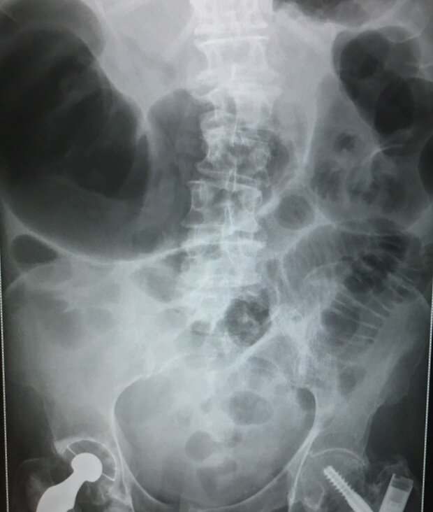
Abdominal X-Ray
AXR erect/supine – 75% sens, 50% spec for SBO
SBO - Valvulae conniventes are visible, predominantly centrally located dilated loops (>3cm) proximal to obstruction, > 5 air/fluid levels
LBO - Haustra visible, Peripherally located dilated loops (>6cm), May be concurrent dilated small bowel, Rectum has little or no air, Pneumoperitoneum
Caecal Volvulus - single gas fluid level in dilated (>9cm) air filled caecum in mid abdomen of LUQ. “Kidney bean sign”
Sigmoid Volvulus - Single dilated loop of colon with both ends orientated towards pelvis. “Coffee bean sign”

Case courtesy of Assoc Prof Frank Gaillard, Radiopaedia.org, rID: 17957
Erect CXR
Looking for free air under diaphragm indicating perforation

Case courtesy of Dr Jeremy Jones, Radiopaedia.org, rID: 6135
CT Abdomen with Contrast
Gold standard. 95% sensitive and specific
Can establish diagnosis, level of obstruction, the cause and presence of complications
Management and Disposition
Initial Resuscitation
IV Access and fluids as clinically indicate
Pt may be in shock and require inotropes and organ support
Specific Treatment
Nil orally.
If vomiting prominent in SBO —> NG on free drainage
If septic consider broad spectrum IV antibiotics that provide gram negative and anaerobic cover as per local guidelines
e.g. IV Co-amoxiclav + IV Metronidazole
Large bowel obstruction/sigmoid volvulus —> decompression with rectal tube by surgeons may be effective
Laparotomy required if concern for ischaemia, perforation, peritonitis etc.
Symptomatic Treatment
Analgesia as required asap – usually IV opioid
Anti-emetic as required
Disposition
Refer all suspected cases to the on call surgical team for admission.
Conservative watch and wait vs operative management is a decision for the surgeons
References
1. Dunn et al. Chapter 37: Abdominal pain. Bowel Obstruction. “The Emergency Medicine Manual” 5th Edition. Vol 1.
This blog was written by Dr. Deirdre Glynn and was last updated in October 2020