
Haemorrhagic Shock
Introduction
Haemorrhagic shock is a type of hypovolaemic shock that occurs due to massive haemorrhage resulting in inadequate organ perfusion and tissue oxygenation. Common causes of haemorrhagic shock include major trauma, intra or postoperative haemorrhage, obstetric haemorrhage, aneurysm rupture and ectopic pregnancy rupture.
Massive haemorrhage is defined as one of;
Acute transfusion of 4 or more units of blood in 1 hr with ongoing haemorrhage
Loss of > 50% of blood volume in 3 hrs or less
Loss of 1 or more blood volumes within 24 hrs (5L blood in 70kg adult)
In the setting of trauma it is convenient to consider the potential source of haemorrhage using the mnemonic “On the Floor and Four More”
Floor – Major external wound e.g. scalp, arterial injury to a limb
Chest - e.g. massive haemothorax, great vessel injury
Abdomen – e.g. Liver, spleen laceration, vessel injury
Pelvis/Retroperitoneum – e.g. pelvic fracture, kidney laceration
Long Bone Fracture – especially femur fracture
Clinical Features
Symptoms
Pain
suggesting source of bleeding e.g. abdomen, long bone, shoulder tip,
Respiratory
Shortness of Breath
Feeling cold, thirsty, wanting to change position constantly, urgent desire to open bowels
Signs
Signs of Shock
loss of airway, reduced GCS, hypoxia, tachypnoea, hypotension, tachycardia, pallor, reduced cap refill
Skin
bruising indicating source of bleeding, mottling
Resp
Signs of Massive haemothorax e.g decreased a/e, dull to percussion, tenderness
Abdomen
Signs of haemoperitoneum e.g. distension, tenderness, bruising, guarding
Pelvis
Genital bruising, blood at urinary meatus
Long Bone
compartment swelling, bruising, rotated limbs
Differential Diagnosis
Other causes of shock
Obstructive shock (2nd most common cause of shock in trauma)
e.g. Cardiac Tamponade, tension pneumothorax, massive PE or fat embolism
e.g. myocardial contusion, secondary to severe traumatic brain injury
Neurogenic shock
secondary to traumatic spinal cord injury
Other causes of SIRS and vasodilatory or distributive shock
e.g. Pancreatitis, Burns, Toxicology
Hypovolaemic shock secondary to dehydration
Clinical Investigations

Bedside
VBG
Low pH, low base excess + high lactate are all indicators of haemorrhagic shock.
Haemoglobin has no diagnostic value in the early phase of massive haemorrhage as haemodilution as not occurred.
Monitor ionized calcium on VBG
eFAST
can identify source of bleeding (chest/abdomen).
Can out rule other causes of shock e.g. tension pneumothorax, tamponade
ECG - ? ST elevation

Laboratory
FBC – Monitor Hb trend. Platelets very important for clotting
Group and Cross Match at least 6 units
Coag – Assess for & monitor development of coagulopathy
U&E, LFT, Amylase - baseline trauma bloods

Radiology
Portable CXR and Pelvic XR in resus post primary survey in the unstable patient
If patient stable CT as clinically indicated to identify injuries and guide further management
Plain films as clinically indicated in stable patient
Management & Disposition
Initial Resuscitation
Call for Help. Pt is critically unwell and will require lots of people to manage. Trauma patient should be managed by Trauma Team including (not limited to) ED, Surgeons and Anaesthesia/ICU
Declare “Massive Transfusion/Code Red”
Multiple (at least 2) wide bore access
Only fluid to be administered to ED patients with haemorrhagic shock is Blood Products
In the immediate setting = O negative Packed Red Blood Cells
Specific Treatment
Single most important treatment is Control & Stop the Bleeding
For internal haemorrhage = may involve urgent transfer to theatre or Interventional Radiology
Other ways to stop bleeding include pelvic binder, splinting long bones, direct pressure to bleeding wounds, application of tourniquets.
Damage Control Resuscitation (3 steps)
Permissive Hypotension = Aiming for a BP that is high enough to adequately perfuse organs but low enough to reduce risk of dilutional coagulopathy and clot disruption. MAP of approx 65mmHg
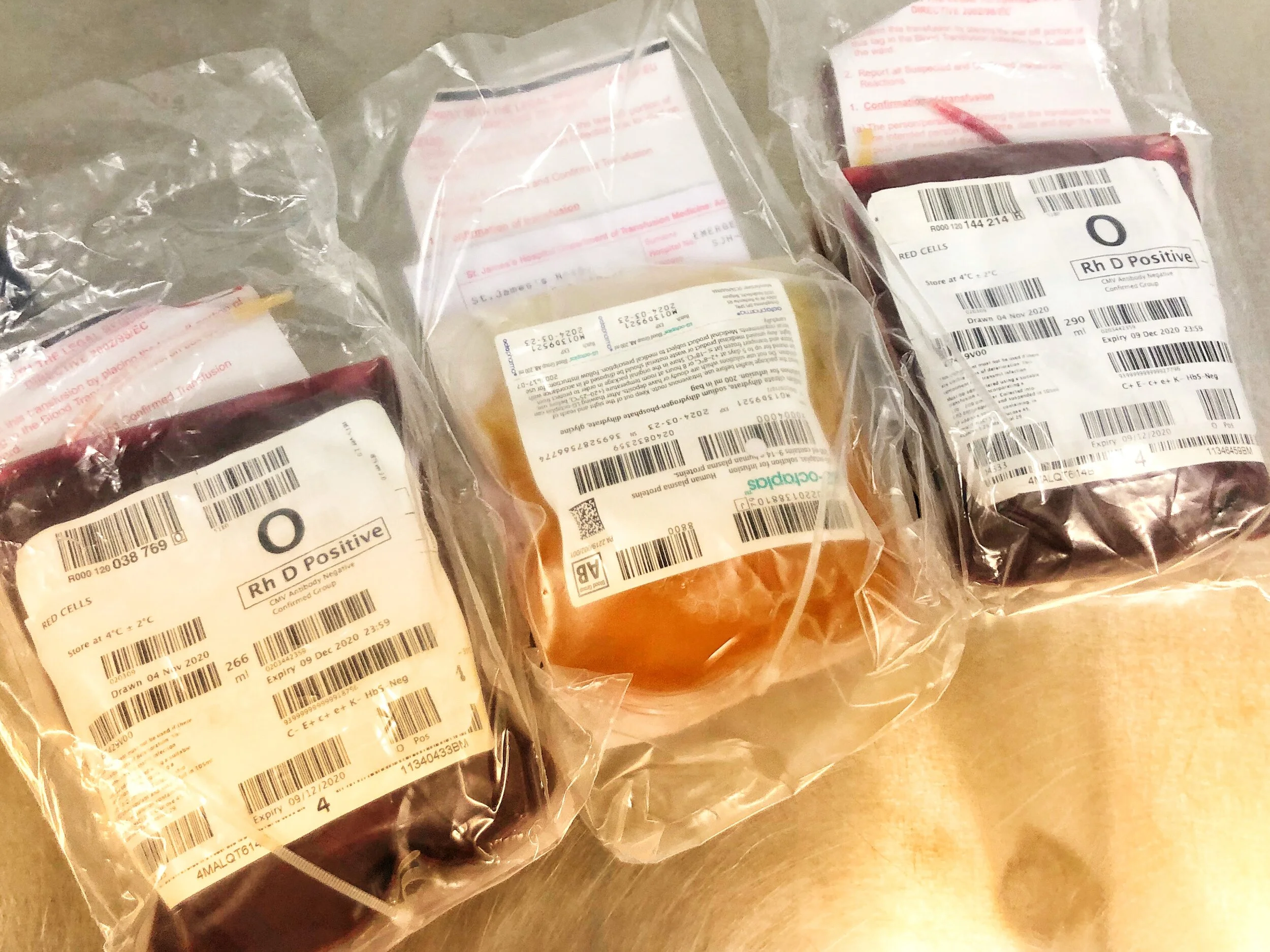
Haemostatic Resuscitation = Transfuse blood products (Red Cells + Plasma + Platelets) at ratio of 1:1:1.
Do not give crystalloid due to risk of causing coagulopathy
Damage Control Surgery e.g. abdominal or pelvic packing to stop bleeding asap.
Avoid the Lethal Triad of;
1. Hypothermia
2. Acidosis
3. Coagulopathy
IV Tranexamic Acid 1g bolus and 1g infusion within 3 hours of trauma
Disposition
Once bleeding has been controlled (either in ED, theatre or radiology) the patient should be admitted to ICU for ongoing coagulopathy treatment and organ support as required.
References
1. Massive Blood Loss: Blood and Blood Product Replacement Guideline. SJH. LabMed Directorate, Department of Transfusion Medicine
2. Managing Major Haemorrhage in the Emergency Department. Rcemlearning.co.uk
3. Holcomb JB et al. The PROPPR randomized clinical trial. JAMA 2015
This blog was written by Dr Deirdre Glynn and was last updated in November 2020